Introduction: How Did Living Organisms Learn to Perceive, Transmit and Process Information?
Long before human beings developed machines capable of performing calculations, recognising patterns, interpreting language or generating complex lines of reasoning, nature had already devised an extraordinarily sophisticated information-processing system: the nervous system.
Emerging during the evolutionary history of animals, it represents one of the most decisive biological innovations in the history of life. It made rapid coordination between different parts of an organism possible, along with the perception of its environment, the adaptation of its behaviour and, progressively, the emergence of increasingly sophisticated capacities for integration.
The nervous system, however, is not simply an accumulation of cells responsible for transmitting electrical impulses. It is a dynamic biological architecture involving electrical, chemical, molecular and metabolic mechanisms. Its fundamental units, neurons, communicate through networks of connections known as synapses, while other cells, collectively referred to as glial cells, play an active part in their functioning.
This organisation enables living organisms to transform physical or chemical information into appropriate responses. A change in temperature, mechanical pressure, a variation in light intensity or the presence of a substance in the environment can therefore trigger a succession of cellular events that ultimately produce a behavioural, muscular, glandular or physiological response.
The history of the nervous system is also the story of the progressive transformation of biological architectures. From diffuse networks, without a clearly defined control centre, to the ganglionic organisation of invertebrates and, subsequently, the centralised nervous systems of vertebrates, evolution has explored several ways of coordinating the activities of an organism.
In human beings, this history has culminated in a particularly highly developed system, capable of processing sensory information simultaneously, regulating vital functions, producing complex movements and supporting cognitive activities such as memory, language, learning and abstract representation.
This biological organisation has also profoundly influenced another scientific field: artificial intelligence.
Artificial neural networks, which today form one of the foundations of machine learning and modern artificial intelligence, explicitly draw on certain general properties of biological networks. They use interconnected computational units, adjustable parameters and learning mechanisms designed to extract patterns from data.
They are not, however, faithful reproductions of the brain. Biological neurons are living cells: electrochemical, metabolically active and deeply integrated into their environment. Artificial neurons, by contrast, are mathematical constructs executed on computer architectures.
Understanding this distinction requires a return to the foundations of neurobiology.
To grasp the connections between the brain and artificial intelligence, we must first understand how the nervous system emerged, how neurons became specialised, how they communicate and how their interactions give rise to the collective properties of living organisms.
1. The Origins of the Nervous System: When Evolution Invented Biological Communication
1.1. Before Neurons: The Earliest Forms of Perception and Response
The ability to respond to one’s environment is far older than the nervous system.
Single-celled organisms, which appeared long before multicellular animals, already possess various mechanisms that enable them to detect changes in their surroundings and respond to them.
A cell can, in particular, react to light, temperature, the concentration of certain molecules or changes in its chemical environment. Such responses involve membrane proteins, molecular receptors, intracellular signalling cascades and regulatory mechanisms.
Some unicellular species also possess structures specialised in perception or movement.
In certain euglenas, for example, a photosensitive organelle helps orient movement in response to light. Cilia and flagella, meanwhile, enable organisms to move or change their position.
These mechanisms do not constitute a nervous system. Nevertheless, they demonstrate that several fundamental principles of biological communication were already present before neurons appeared.
The detection of a signal, its conversion into a cellular response and the coordination of different functions are all ancient properties of living systems.
The emergence of the nervous system therefore did not mark the sudden birth of sensitivity or responsiveness. Rather, it represented a new way of organising these properties at the level of a multicellular organism.
1.2. Multicellular Organisms and the Need to Coordinate Functions
The emergence of multicellular organisms profoundly transformed biological constraints.
In an organism consisting of a single cell, its various metabolic functions are brought together within the same cellular space. In a multicellular organism, by contrast, cells gradually become specialised.
Some take part in digestion, others in protection, reproduction, contraction or exchanges with the environment.
This specialisation creates a new problem: how can different cells, sometimes located far apart, be coordinated so that they contribute to a common response?
Multicellular organisms have developed several communication systems.
Chemical signalling, in particular, enables cells to transmit information through molecules that diffuse into their surroundings or are transported by internal fluids.
Hormonal communication is one of the most sophisticated forms of this organisation. It makes it possible to regulate functions that may be located far from the site where the signal is produced.
The nervous system offers another solution: transmitting information rapidly through specialised cells capable of receiving, transforming and propagating electrical and chemical signals.
This ability to organise rapid and relatively precise communication between different regions of the body represented a considerable advantage for mobile animals.
An organism capable of rapidly detecting a predator, changing direction, coordinating its muscles and adapting its behaviour may have better chances of survival than one whose responses are slower or less coordinated.
The nervous system should therefore be understood as an innovation in biological coordination.
1.3. Sponges: An Essential Example for Understanding Evolution
Sponges, or Porifera, occupy a particularly interesting position in the study of the evolution of nervous systems.
They are multicellular animals that possess neither neurons nor an organised nervous system in the conventional sense.
Their functioning nevertheless relies on remarkable mechanisms of cellular communication.
Their organisation notably involves cells specialised in water circulation, the capture of food particles, exchanges with the environment and the regulation of the organism’s activities.
Sponges can modify their functioning in response to various environmental stimuli. They also possess molecular communication mechanisms between their cells.
Their existence demonstrates that an animal can perform coordinated biological functions without possessing a brain or a neuronal network.
It would therefore be incorrect to regard the nervous system as a universal prerequisite for animal life.
Evolution has produced different strategies for biological organisation, and some lineages have retained architectures without neurons.
Placozoans, small marine animals with a relatively simple organisation, provide another important example of this diversity.
1.4. The First Neural Networks: The Diffuse Organisation of Cnidarians
A true nervous organisation appears among the Cnidarians, a group that includes jellyfish, sea anemones and corals.
Unlike the centralised nervous system of vertebrates, their architecture is based primarily on a diffuse network of nerve cells.
Neurons are distributed across different regions of the organism and communicate through connections that coordinate responses to stimuli.
This organisation is particularly well suited to animals whose bodies do not necessarily have a dominant direction of movement.
In a jellyfish, for example, information originating in different parts of the body can contribute to coordinating the contractions responsible for locomotion.
Some species also possess specialised sensory structures.
Ocelli detect certain forms of light-related information, while statocysts contribute to the perception of orientation and gravity.
The significance of this organisation lies in its ability to produce functional coordination without requiring a centralised brain.
It is also interesting to note that animals with diffuse neural networks can display elementary forms of learning, notably habituation and sensitisation.
Habituation is the gradual reduction of a response to a repeated stimulus that has no particular consequence.
Sensitisation, by contrast, refers to an increase in responsiveness following a sufficiently significant stimulus.
These phenomena demonstrate that certain fundamental properties of learning do not necessarily require a brain comparable to that of vertebrates.
1.5. Ctenophores: An Evolutionary History That Remains Scientifically Debated
Ctenophores, also known as comb jellies, constitute a particularly interesting group for understanding the origins of nervous systems.
These marine animals possess a diffuse neural network, sensory cells and specialised coordination structures.
Their position in the evolutionary tree of animals is, however, the subject of considerable scientific debate.
Contemporary research is particularly concerned with the origin of ctenophore neurons and the possibility that certain components of their nervous system may have evolved independently of those found in other major animal groups.
Two broad hypotheses are being investigated:
- A common origin of nervous systems, followed by substantial diversification and transformation across different animal lineages.
- Several evolutionary events that led to the emergence of nervous systems displaying certain functional similarities through convergence.
The difficulty lies, in particular, in the fact that living organisms are not unchanged representatives of their ancestors.
Every lineage has continued to evolve since diverging from other groups.
It is therefore difficult to reconstruct the precise characteristics of the first animal nervous system by observing contemporary species alone.
This question remains an active field of research in evolutionary biology.
One essential finding is that the nervous system should not be viewed as an architecture that appeared once and for all and was subsequently merely perfected. Its history involves divergence, specialisation and, probably, processes of evolutionary convergence.
For further insight into this question, the work of Maria Y. Sachkova, published in 2024 in the journal Evolution & Development, provides a particularly valuable reference.
1.6. Bilateral Symmetry and the Emergence of Neural Centralisation
An important transformation appeared with the diversification of animals displaying bilateral symmetry.
These organisms generally possess an anterior and a posterior axis, as well as two relatively symmetrical lateral sides.
This anatomical organisation favours the development of directional behaviour.
An animal that habitually moves forwards encounters its environment first and foremost with its anterior region.
Sensory organs therefore tend to become concentrated in this part of the body.
This sensory concentration is frequently accompanied by a concentration of neural structures.
This phenomenon, known as cephalisation, refers to the progressive organisation of an anterior region specialised in integrating certain types of information and coordinating responses.
In flatworms, particularly planarians, one can observe an organisation combining anterior ganglia, longitudinal nerve cords and transverse connections.
The nervous system thus begins to display a more hierarchical architecture.
Sensory information can be channelled towards regions where neurons are concentrated, before contributing to the coordination of movements.
This organisation does not yet correspond to a brain comparable to that of vertebrates, but it marks an important stage in the history of neural centralisation.
1.7. Annelids: The Specialisation of Ganglia and Neural Circuits
In annelids, such as earthworms, the nervous system displays a more structured organisation.
It notably includes an anterior neural region and a ventral nerve cord comprising segmental ganglia.
Each ganglion contributes to processing information and coordinating the activities of the corresponding body region.
This organisation makes it possible to combine a degree of local autonomy with the overall coordination of the organism.
A segment can thus contribute to motor responses while remaining integrated into a broader set of neural circuits.
This architecture provides an interesting example of distributed information processing.
It also presents a functional analogy with certain computer systems in which several specialised units operate simultaneously while exchanging information.
This organisational resemblance should not, however, be confused with an identity of functioning.
Biological ganglia consist of living cells capable of modifying their activity, their connections and their molecular environment.
1.8. Arthropods: The Sophistication of Invertebrate Neural Networks
Arthropods, which include insects, spiders and crustaceans, possess particularly diverse nervous systems.
Their organisation generally relies on an anterior neural region and a ventral nerve cord containing several ganglia.
In some species, the ganglia of several segments progressively fuse, giving rise to more concentrated neural centres.
Insects, in particular, possess specialised brain structures involved in sensory integration, orientation, locomotion and learning.
The optic lobes process visual information, while the mushroom bodies contribute to various mechanisms of learning and memory.
The fruit fly, Drosophila, is a major model organism in this respect for neuroscience.
Its nervous system, far less extensive than that of a mammal, makes it possible to investigate experimentally the relationships between neural circuits, cellular activity and behaviour.
Research on insects shows that a relatively compact neural architecture can produce complex behaviours.
The number of neurons alone is therefore insufficient to determine the functional capabilities of a nervous system.
The organisation of circuits, cell specialisation, learning mechanisms and interactions with the environment also play a decisive role.
1.9. Molluscs: Remarkably Different Neural Architectures
Molluscs display a remarkable diversity of nervous systems.
In relatively sedentary species, neural structures may remain fairly dispersed and organised around specialised ganglia.
In cephalopods, by contrast, neural organisation reaches an exceptional degree of sophistication.
Octopuses, squids and cuttlefish possess highly developed brains, complex sensory systems and remarkable behavioural abilities.
Their organisation nevertheless relies on a distinctive distribution of functions between the central brain and peripheral circuits.
In the octopus, a substantial proportion of neural processing is associated with the arms and their local circuits.
The arms contain numerous neurons and can perform complex motor operations without every movement having to be specified in full by the central brain.
This organisation illustrates another way of distributing information-processing capabilities throughout an organism.
Cephalopods thus provide a remarkable example of evolutionary convergence with vertebrates.
Very distantly related animal lineages have independently developed certain complex sensory and behavioural capacities.
1.10. Vertebrates: Centralisation Around a Dorsal Nervous System
Vertebrates display an organisation characterised by a dorsal central nervous system.
This develops from an embryonic neural tube, which gives rise to the brain in its anterior region and the spinal cord in its posterior region.
This organisation is one of the fundamental characteristics of the vertebrate body plan.
The central nervous system is protected by specialised anatomical structures:
- The skull protects the brain.
- The vertebral column protects the spinal cord.
- The meninges envelop the central nervous system.
- Cerebrospinal fluid contributes to its mechanical protection and physiological environment.
The evolution of vertebrates subsequently produced highly diverse nervous systems.
Fish, amphibians, reptiles, birds and mammals display brain architectures adapted to their ecological, sensory and behavioural constraints.
This should not be understood as a linear progression in which each group is necessarily more evolved than the one preceding it.
Each lineage represents a particular evolutionary solution.
The human nervous system is one such solution, characterised notably by the extensive development of cortical structures and association networks.
2. The Neuron: The Fundamental Cell of Neural Communication
2.1. A Cell Specialised in Information Processing
A neuron is a living cell specialised in receiving, integrating and transmitting signals.
Its distinctive features lie in its morphological and physiological organisation.
Unlike a cell whose activity is primarily limited to local metabolic exchanges, a neuron can receive information from several regions, integrate it and produce an electrical or chemical response that can be transmitted to other cells.
This specialisation rests on two essential properties:
Excitability refers to the ability of the cell membrane to respond to a stimulus by changing its electrical state.
Conductivity refers to the ability to propagate an electrical signal along certain regions of the cell.
These two properties enable neurons to participate in extraordinarily sophisticated communication circuits.
There is, however, considerable diversity among neurons.
Some transmit sensory information originating in the skin or internal organs.
Others control muscle contractions.
Still others ensure communication between different regions of the central nervous system.
The neuron is therefore not a uniform unit. Hundreds of neuronal categories exist, differentiated by their morphology, function, gene expression and electrophysiological properties.
2.2. The History of the Discovery of the Neuron: From Reticular Theory to the Neuron Doctrine
Our understanding of the neuron was profoundly transformed by advances in microscopy and histology.
During the nineteenth century, researchers sought to understand how nervous tissue was organised.
A major controversy emerged between two competing conceptions.
Reticular theory, notably defended by Camillo Golgi, held that nervous tissue formed a continuous network in which cellular extensions were directly connected.
A different conception, progressively championed by Santiago Ramón y Cajal, maintained instead that the nervous system was composed of distinct cells that established functional relationships through specialised contacts.
Cajal’s work, made possible in particular by the staining techniques developed by Golgi, profoundly transformed neuroanatomy.
The neuron doctrine became one of the foundations of modern neuroscience.
Advances in electron microscopy subsequently made it possible to observe directly the specialised structures separating two nerve cells: synapses.
This discovery confirmed that neuronal communication generally relies on functional contacts between distinct cells, rather than on widespread cytoplasmic continuity.
The historical controversy is also relevant to artificial intelligence: understanding the brain means understanding not only the units of which it is composed, but above all the relationships established between them.
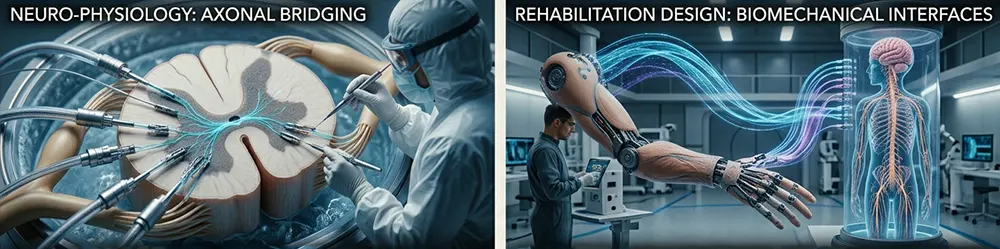
2.3. The Four Main Functional Regions of a Neuron
A neuron can be described in terms of several fundamental anatomical components.
The cell body: the neuron’s metabolic centre
The cell body, also known as the soma or perikaryon, contains the nucleus and most of the organelles required for the cell to function.
It is responsible, in particular, for protein synthesis, energy production and the maintenance of cellular structures.
It contains:
- The nucleus, which holds the genetic information.
- Mitochondria, which contribute to energy production.
- The endoplasmic reticulum, involved in protein synthesis.
- The Golgi apparatus, which contributes to protein maturation and transport.
- Ribosomes, which are required for protein production.
- The cytoskeleton, which maintains cellular organisation and intracellular transport.
Nissl bodies, visible with certain histological stains, correspond primarily to regions rich in rough endoplasmic reticulum and ribosomes.
They reflect the neuron’s considerable protein-synthesis activity.
The cell body is therefore not merely an anatomical support structure. It is a metabolic centre essential to maintaining neuronal extensions.
Dendrites: The Receiving Structures
Dendrites are generally branched cellular extensions.
They form an important part of the neuron’s receptive surface.
They receive information transmitted by other cells at synapses.
Their tree-like shape makes it possible to multiply the opportunities for connection.
Some neurons possess particularly extensive dendritic arbors.
Pyramidal neurons in the cerebral cortex, for example, can receive a considerable number of connections originating in different regions of the brain.
Dendrites are not, however, merely passive cables.
They possess electrical properties that enable them to modify and locally transform the signals they receive.
Some can also generate specific electrical events.
Dendritic spines are small structures that increase the surface area available for synaptic contacts.
They play an important role in synaptic plasticity, that is, in the lasting modification of certain neuronal connections.
The Axon: The Pathway Along Which Signals Travel
The axon is generally a single extension, sometimes extremely long, that propagates action potentials towards other cells.
Its length varies considerably depending on the type of neuron.
Some axons are very short and participate in local circuits.
Others can extend across a substantial part of the organism.
The axons of motor neurons responsible for controlling muscles, for example, can extend from the spinal cord to the muscles of the limbs.
The axon has a specialised membrane known as the axolemma and an internal cytoplasm called the axoplasm.
It contains mitochondria and cytoskeletal structures, notably microtubules and neurofilaments.
These components help maintain its structure and transport molecules between the cell body and the nerve terminals.
Axon terminals: transmitting information to another cell
At its extremity, the axon may branch to form a terminal arborisation.
These branches end in synaptic boutons.
The boutons contain, among other structures, mitochondria and synaptic vesicles.
The latter contain neurotransmitters, which are released when the electrical signal reaches the axon terminal.
Transmission can then take place to another neuron, a muscle cell or a glandular cell.
2.4. The Different Shapes of Neurons
Neuronal morphology varies considerably according to function.
Several broad categories can be distinguished.
Unipolar or pseudounipolar neurons
They possess a main extension that divides into two branches.
They are found, in particular, in the sensory ganglia of vertebrates.
One branch extends towards peripheral receptors, while the other connects to the central nervous system.
Bipolar neurons
They possess two main extensions, generally located on opposite sides of the cell body.
They are found, in particular, in certain sensory structures, such as the retina and the olfactory pathways.
Multipolar neurons
They possess several dendrites and one axon.
They form a widespread category within the central nervous system.
Motor neurons and many cortical neurons belong to this category.
The morphological diversity of neurons reflects their functional specialisation.
A sensory neuron, a motor neuron and a neuron involved in memory do not necessarily share the same electrical properties, connections or molecular organisation.
3. Myelin: The Biological Insulation That Speeds Up Neural Transmission
3.1. Why Do Axons Need a Sheath?
The propagation of an electrical signal along an axon requires a particular membrane organisation.
Some axons are surrounded by a substance rich in lipids and proteins known as myelin.
This sheath acts as an electrical insulator and profoundly modifies the way in which a signal propagates.
Myelin notably increases transmission speed and reduces the ionic exchanges required for the propagation of the action potential.
It therefore represents a particularly important adaptation in nervous systems containing long communication pathways.
3.2. The Cells Responsible for Producing Myelin
Myelin is produced by two major categories of cells, depending on the anatomical location.
In the peripheral nervous system, it is formed by Schwann cells.
In the central nervous system, it is produced by oligodendrocytes.
This distinction is important.
A myelinating Schwann cell generally surrounds one segment of a single axon.
An oligodendrocyte, by contrast, can produce several myelin segments on different axons.
Myelin is therefore not a uniform sheath deposited independently of nervous tissue.
It results from a close interaction between axons and glial cells.
3.3. Nodes of Ranvier and Saltatory Conduction
The myelin sheath contains regular interruptions known as nodes of Ranvier.
These regions allow significant ionic exchanges between the inside and outside of the axon.
In a myelinated fibre, the action potential is regenerated at these nodes.
The signal therefore appears to travel in successive leaps from one node to the next.
This phenomenon is known as saltatory conduction.
It enables rapid propagation and is energetically more efficient than conduction in a non-myelinated fibre with comparable characteristics.
Conduction speed depends notably on axon diameter, myelin thickness and the organisation of myelinated segments.
Some nerve fibres can therefore transmit signals at speeds well above several dozen metres per second.
3.4. Myelin and Neurological Diseases
Myelin plays a decisive role in the normal functioning of the nervous system.
Damage to this sheath can disrupt the transmission of electrical signals.
Multiple sclerosis is one of the best-known demyelinating diseases of the central nervous system.
It involves inflammatory and immune-mediated damage to myelin, which can interfere with neural conduction.
Depending on the regions affected, the consequences may involve motor function, sensation, vision or other neurological functions.
This condition illustrates the importance of the cellular environment and supporting structures in the functioning of neurons.
4. The Electrical Functioning of the Neuron: How Does a Nerve Signal Arise?
4.1. The Neuronal Membrane and the Resting Potential
The electrical functioning of a neuron is based on the properties of its plasma membrane.
This membrane separates two environments with different ionic compositions.
The intracellular environment contains, in particular, a high concentration of potassium, whereas the extracellular environment is relatively rich in sodium.
These differences in concentration contribute to the creation of an electrical potential difference between the inside and the outside of the cell.
At rest, the inside of the neuron generally has a negative potential relative to the extracellular environment.
This difference in electrical potential is known as the resting potential.
It frequently lies between approximately -60 and -70 millivolts, although values vary according to cell type and physiological conditions.
Several mechanisms contribute to maintaining it:
- Ion concentration gradients.
- The selective permeability of the membrane.
- Ion channels.
- The activity of the sodium-potassium pump.
The sodium-potassium pump uses the energy supplied by ATP to transport three sodium ions out of the cell and two potassium ions into it.
It therefore contributes to maintaining the ionic gradients required for electrical activity.
It is important to point out that the resting potential is not produced solely by this pump: the properties of membrane permeability, particularly with regard to potassium, also play an essential role.
4.2. The Action Potential: A Fundamental Electrical Impulse
When a neuron receives stimulation, its membrane potential may change.
If the stimulation produces sufficient depolarisation to reach a threshold, an action potential may be generated.
An action potential is a brief electrical event characterised by a rapid change in membrane potential.
Its course comprises several stages.
First stage: depolarisation
The opening of voltage-gated sodium channels causes a rapid influx of sodium ions into the cell.
The membrane potential gradually becomes less negative and may even become temporarily positive.
Second stage: repolarisation
Sodium channels become inactivated, while voltage-gated potassium channels contribute to the outward movement of potassium ions.
The membrane potential gradually returns towards its initial value.
Third stage: transient hyperpolarisation
In some neurons, the delayed closure of potassium channels produces a temporary hyperpolarisation.
The membrane then becomes momentarily more negative than its resting potential.
Fourth stage: return to the resting state
Ionic regulatory mechanisms and the properties of membrane channels make it possible to restore the conditions required for renewed electrical activity.
4.3. The All-or-None Principle
The action potential follows a fundamental principle: the all-or-none law.
Once the excitation threshold has been reached, the action potential is triggered with a relatively stereotyped amplitude.
A stronger stimulus therefore does not necessarily produce an action potential of greater amplitude.
Information about the intensity of a stimulus is represented, among other things, by the frequency of action potentials, their temporal organisation and the coordinated activity of neuronal populations.
A more intense stimulus may thus cause an increase in firing frequency or recruit a greater number of neurons.
This distinction is essential to understanding how neural networks function.
The neuron does not simply transmit an electrical value proportional to the intensity of the stimulus. It transforms the information it receives into structured temporal activity.
4.4. Refractory Periods
After producing an action potential, a neuron passes through a period during which it cannot immediately generate another one under the same conditions.
Two phases can be distinguished:
- The absolute refractory period, during which a new action potential cannot be triggered.
- The relative refractory period, during which a stronger-than-usual stimulus may be required.
These mechanisms help limit the maximum firing frequency and organise the propagation of signals along the axon.
4.5. Signal Propagation Along the Axon
The action potential propagates through local electrical currents that modify the state of the membrane adjacent to the region that has already been activated.
In non-myelinated fibres, this propagation takes place progressively along the membrane.
In myelinated fibres, saltatory conduction enables faster propagation between the nodes of Ranvier.
Nevertheless, it is important to distinguish between the propagation of an action potential and synaptic transmission.
The former is an electrical event travelling along a single cell.
The latter is communication between two distinct cells.
This distinction is one of the foundations of neuronal organisation.
5. The Synapse: The Language of Communication Between Neurons
5.1. An Interface Between Two Cells
A synapse is a specialised communication site between two cells.
In the most familiar arrangement, it links the axon terminal of a presynaptic neuron to a receptive region of another neuron.
Three components can then be distinguished:
- The presynaptic compartment, which transmits the signal.
- The synaptic cleft, which separates the two membranes.
- The postsynaptic compartment, which receives the message.
The synapse is an essential structure for understanding how the brain works.
It enables not only the transmission of information, but also its modulation.
Depending on the properties of the connection and the state of the network, the incoming signal may be amplified, attenuated, transformed or inhibited.
5.2. Chemical Transmission
Most synapses in the central nervous system communicate through neurotransmitters.
The general mechanism unfolds in several stages:
- An action potential reaches the axon terminal.
- Depolarisation causes voltage-gated calcium channels to open.
- Calcium ions enter the presynaptic terminal.
- This increase in intracellular calcium triggers the fusion of certain synaptic vesicles with the membrane.
- Neurotransmitters are released into the synaptic cleft.
- They bind to specific receptors located on the postsynaptic membrane.
- These receptors modify the electrical or biochemical activity of the receiving cell.
The chemical message is then eliminated, taken back up or transformed through various mechanisms.
Synaptic transmission therefore involves a conversion between two forms of communication: electrical and chemical.
5.3. The Main Neurotransmitters
Neurotransmitters are molecules that enable communication between neurons and other cells.
Their diversity partly explains the functional richness of the nervous system.
Acetylcholine
Acetylcholine is involved, in particular, in neuromuscular transmission.
It enables communication between motor neurons and skeletal muscle fibres.
It also plays a role in certain functions of the autonomic nervous system and in various brain circuits.
At the neuromuscular junction, its release activates nicotinic receptors, triggering an electrical response in the muscle fibre.
Acetylcholine is then rapidly broken down by an enzyme known as acetylcholinesterase.
This breakdown brings the chemical signal to an end.
Glutamate
Glutamate is the main excitatory neurotransmitter in the central nervous system.
It is involved in numerous mechanisms of neuronal communication.
It also plays an essential role in synaptic plasticity and in certain processes of learning and memory.
Its receptors include AMPA, NMDA and several metabotropic receptors.
These different receptors have distinct functional properties.
GABA
Gamma-aminobutyric acid, generally referred to by the abbreviation GABA, is the main inhibitory neurotransmitter in the adult brain.
It is involved in regulating neuronal activity.
By promoting certain inhibitory responses, it helps prevent excessive excitation within networks.
GABA thus contributes to the essential balance between excitation and inhibition.
Glycine
Glycine is also an important inhibitory neurotransmitter.
It is involved, in particular, in the spinal cord and certain regions of the brainstem.
It contributes to the regulation of motor and sensory circuits.
Dopamine
Dopamine plays an essential role in several brain circuits.
It is involved, in particular, in:
- Motivation.
- Reinforcement learning.
- The selection of certain actions.
- The regulation of movement.
- Reward mechanisms.
Dopamine should not be reduced to a simple pleasure molecule.
Its functioning depends on the circuits in which it operates, the receptors involved and the dynamics of its release.
Serotonin
Serotonin is involved in numerous regulatory mechanisms.
It contributes, in particular, to the control of mood, sleep, appetite, pain and various behavioural processes.
Its action is particularly complex because of the diversity of its receptors.
Noradrenaline
Noradrenaline is involved in alertness, attention, the stress response and the modulation of brain activity.
It contributes, in particular, to adapting the functioning of the brain to the demands of its environment.
5.4. Excitatory and Inhibitory Synapses
Not all synapses produce the same effect.
Some promote the activation of the postsynaptic neuron.
Others reduce its probability of generating an action potential.
Excitatory and inhibitory synapses can therefore be distinguished.
An excitatory synapse generally produces an excitatory postsynaptic potential.
An inhibitory synapse produces an inhibitory postsynaptic potential or inhibition through shunting, depending on the ion channels involved.
This distinction is fundamental.
A functioning brain does not rely on the permanent excitation of neurons, but on a dynamic balance between different mechanisms of activation and regulation.
Inhibition notably makes it possible to limit certain responses, select information and stabilise networks.
5.5. The Different Types of Synapses
Neuronal communication can take several anatomical forms.
Axo-dendritic synapses connect an axon to a dendrite.
Axo-somatic synapses establish contact with the cell body.
Axo-axonic synapses act on another axon and may modulate presynaptic transmission.
Synapses also exist between neurons and effector cells.
The neuromuscular junction is the classic example of a connection between a motor neuron and a muscle fibre.
5.6. Electrical Synapses: Communication Without Conventional Neurotransmitters
Not all synapses rely on the release of neurotransmitters.
Some are electrical.
They use communicating junctions, known as gap junctions, which allow ionic currents to pass directly between two cells.
This organisation generally permits very rapid transmission.
It can also help synchronise the activity of several neurons.
Electrical synapses are present in different animal nervous systems, including those of vertebrates.
They demonstrate that neuronal communication can rely on several complementary mechanisms.
6. Glial Cells: The Indispensable Partners of Neurons
6.1. Moving Beyond the Traditional View of Supporting Cells
For a long time, glial cells were presented primarily as supporting elements of nervous tissue.
This representation is now inadequate.
Contemporary research shows that glial cells participate directly in numerous mechanisms required for the functioning of the brain.
They are involved in regulating the extracellular environment, energy metabolism, synaptic communication, immune defence and myelin formation.
The nervous system therefore cannot be understood by studying neurons alone.
Nervous tissue as a whole must be considered an integrated cellular environment.
6.2. Astrocytes
Astrocytes are glial cells characterised by their star-shaped appearance and numerous extensions.
They establish close relationships with neurons and blood vessels.
Their functions include, in particular:
- Regulating extracellular ion concentrations.
- Contributing to the energy metabolism of neurons.
- Taking up certain neurotransmitters.
- Regulating the synaptic environment.
- Contributing to the blood-brain barrier.
- Modulating certain forms of neuronal communication.
Astrocytes play an especially important role in regulating extracellular glutamate.
They thus help prevent an excessive accumulation of this neurotransmitter, which could disrupt neuronal functioning.
They also participate in metabolic exchanges between blood vessels and nervous tissue.
Their role therefore extends far beyond a simple nutritional function.
6.3. Oligodendrocytes
Oligodendrocytes are responsible for producing myelin in the central nervous system.
They surround axons with myelinated segments and contribute to the efficiency of neural conduction.
Their function, however, is not limited to electrical insulation.
They also contribute to the metabolic support of axons and help maintain their integrity.
The relationship between neurons and oligodendrocytes is particularly important in long neural pathways.
6.4. Schwann Cells
Schwann cells perform comparable functions in the peripheral nervous system.
They contribute to the myelination of axons and to their maintenance.
They also play an important role in repair mechanisms following certain peripheral nerve injuries.
Unlike the central nervous system, the peripheral nervous system generally has a greater capacity for axonal regeneration, although this remains variable and depends on numerous factors.

6.5. Microglia: The Brain’s Immune System
Microglia constitute a specialised population of resident immune cells in the central nervous system.
They help monitor the brain environment and respond to harmful events.
Their functions notably include:
- Detecting danger signals.
- Removing cellular debris.
- Phagocytosis.
- Regulating certain inflammatory responses.
- Contributing to the maturation and remodelling of neural circuits.
Microglia also participate in certain mechanisms of synaptic remodelling.
They illustrate how closely immune and nervous system functions are interconnected.
6.6. Ependymal Cells
Ependymal cells line the ventricular cavities of the brain and the central canal of the spinal cord.
They contribute to the organisation of the interfaces between nervous tissue and cerebrospinal fluid.
The choroid plexuses, specialised structures associated with the ventricles, are responsible for much of the production of this fluid.
Cerebrospinal fluid contributes to the mechanical protection of the central nervous system and to the maintenance of its physiological environment.
6.7. A New Understanding of Brain Function
Advances in neurobiology have led researchers to regard the brain as a system composed of several interdependent cell populations.
Neurons carry out much of the rapid electrical and synaptic communication.
Glial cells help maintain the conditions required for this activity and also participate directly in its regulation.
This discovery is important for understanding biological intelligence.
The brain is not a network of neurons isolated from one another.
It is a cellular ecosystem in which interactions between neurons, glial cells, blood vessels and the extracellular environment contribute to overall activity.
7. The Human Nervous System: An Architecture Organised at Several Levels
7.1. The Central Nervous System
In human beings, the nervous system is conventionally divided into two main parts: the central nervous system and the peripheral nervous system.
The central nervous system comprises:
- The brain and spinal cord, collectively referred to as the central nervous system’s principal structures.
- The spinal cord.
More precisely, the central nervous system consists of the encephalon and the spinal cord.
It is responsible for receiving, integrating and generating a large proportion of neural information.
The encephalon is protected by the skull.
The spinal cord lies within the vertebral canal.
These two structures are connected by ascending and descending neural pathways.
7.2. The Peripheral Nervous System
The peripheral nervous system comprises nerves, ganglia and nerve endings.
It provides communication between the central nervous system and the rest of the organism.
Sensory information travels towards the neural centres along afferent pathways.
Motor commands are transmitted to muscles and organs through efferent pathways.
This organisation enables continuous communication between the brain, spinal cord and the different regions of the body.
7.3. The Spinal Cord: A Communication Pathway and a Processing Centre
The spinal cord is an essential structure of the central nervous system.
It transmits information between the brain and the body.
It also participates in generating reflex responses.
Its grey matter contains, in particular, the cell bodies of numerous neurons and local circuits.
Its white matter consists primarily of bundles of axons that carry information between different levels of the spinal cord and the brain.
A simple reflex can be generated without conscious intervention by the brain.
When a painful stimulus reaches a peripheral receptor, the information is transmitted to the spinal cord.
Local circuits can then activate motor neurons that command the withdrawal of the affected limb.
This organisation enables a rapid response, while sensory information simultaneously continues along its pathway towards the brain centres.
7.4. The Brainstem: Vital Functions and Communication Pathways
The brainstem connects the spinal cord to the higher brain structures.
It comprises three major regions:
- The medulla oblongata.
- The pons.
- The midbrain.
It contributes to numerous essential functions.
It contains, in particular, circuits involved in breathing, cardiovascular regulation, swallowing, certain reflexes and the control of wakefulness.
The brainstem is also an important passageway for sensory and motor pathways.
Its functioning is essential to coordinating numerous automatic processes.
7.5. The Cerebellum: Coordination, Balance and Motor Learning
The cerebellum plays a fundamental role in coordinating movements.
It receives information from sensory systems, the spinal cord and brain regions involved in motor control, among other sources.
It contributes to comparing intended movements with the results actually produced.
This function makes it possible to adjust movements and correct certain errors.
The cerebellum is also involved in balance, motor precision and various forms of learning.
Its functions are not, however, limited to movement.
Contemporary research also demonstrates its involvement in certain cognitive processes and in the regulation of complex behaviours.
7.6. The Thalamus: A Hub for Brain Information
The thalamus is a central structure of the diencephalon.
It is involved in transmitting and modulating a wide range of sensory and motor information.
It establishes connections, in particular, with different regions of the cerebral cortex.
The thalamus is not merely a passive relay.
It participates in the selection, modulation and temporal organisation of information circulating through brain circuits.
It is also involved in mechanisms of alertness and certain cognitive functions.
7.7. The Hypothalamus: Maintaining Physiological Balance
The hypothalamus plays a fundamental role in regulating the internal environment.
It is involved, in particular, in:
- Body temperature.
- Hunger and satiety.
- Thirst.
- Biological rhythms.
- Sleep.
- Hormonal regulation.
- Physiological responses to stress.
It maintains close connections with the pituitary gland and several structures of the autonomic nervous system.
This organisation makes it possible to integrate neural and endocrine mechanisms.
The hypothalamus thus illustrates a fundamental property of living organisms: the need to maintain certain physiological constants despite changes in the environment.
This regulation is known as homeostasis.
7.8. The Basal Ganglia: The Selection and Organisation of Movement
The basal ganglia are a group of deep brain structures involved in regulating movement and certain cognitive processes.
They participate, in particular, in action selection, reinforcement learning and the organisation of behaviour.
Circuits involving the striatum, globus pallidus, substantia nigra and subthalamic nucleus play an essential role in this regulation.
Dopamine is involved in several of these circuits.
Parkinson’s disease illustrates the importance of these mechanisms.
The degeneration of dopaminergic neurons in the substantia nigra disrupts the functioning of basal ganglia circuits and can lead to characteristic motor disorders.
7.9. The Cerebral Cortex: Information Integration and Higher Functions
The cerebral cortex is the outer layer of grey matter covering the cerebral hemispheres.
It is involved in numerous complex functions.
Several functional territories can be distinguished.
The frontal cortex
It contributes to planning, action control, decision-making, behavioural regulation and certain language functions.
The parietal cortex
It is involved in integrating sensory information, spatial representation and body perception.
The temporal cortex
It contributes, in particular, to hearing, memory and various aspects of language comprehension.
The occipital cortex
It is a major region involved in processing visual information.
It would nevertheless be reductive to assign each mental function to a single, isolated brain region.
Complex cognitive functions rely on distributed networks linking several brain regions.
Language, memory, attention and decision-making recruit circuits that may extend across different structures.
The cerebral cortex should therefore be understood as a dynamic organisation of interconnected networks.
8. The Embryology of the Nervous System: How Does a Brain Develop?
8.1. The Embryonic Origin of the Nervous System
The human nervous system develops primarily from the ectoderm, one of the three fundamental embryonic germ layers.
This origin is an important characteristic of vertebrate development.
During embryogenesis, certain regions of the ectoderm gradually become specialised to form neural structures.
This process is known as neurulation.
It leads to the formation of the neural tube, from which the brain and spinal cord develop.
8.2. The Neural Plate and the Closure of the Neural Tube
At the beginning of embryonic development, molecular signals originating, in particular, from the underlying axial tissues induce the specialisation of the dorsal ectoderm.
This region thickens to form the neural plate.
The neural plate gradually changes shape.
Its central region sinks inwards, while its edges rise and move towards one another.
The resulting structure is known as the neural groove.
The edges eventually meet and fuse to form the neural tube.
This closure process follows a complex sequence, with differences between embryonic regions.
The anterior part will give rise to the brain.
The posterior part will form the spinal cord.
8.3. Neural Crest Cells: Migrating Cells with Multiple Functions
During neurulation, certain cells located at the boundaries of the neural plate give rise to the neural crest.
These cells possess a remarkable property: they can migrate to different regions of the embryo.
They contribute to the formation of numerous structures.
Their derivatives include, in particular:
- Sensory ganglia.
- Several ganglia of the autonomic nervous system.
- Schwann cells.
- Chromaffin cells of the adrenal medulla.
- Certain components of craniofacial structures.
- Melanocytes.
The neural crest is therefore a remarkable example of cell differentiation and migration during development.
8.4. The First Brain Vesicles
The anterior part of the neural tube gradually expands.
It gives rise to three primary brain vesicles:
- The prosencephalon, or forebrain.
- The mesencephalon, or midbrain.
- The rhombencephalon, or hindbrain.
These structures subsequently undergo further differentiation.
The prosencephalon gives rise to the telencephalon and diencephalon.
The mesencephalon retains its anatomical identity.
The rhombencephalon divides into the metencephalon and myelencephalon.
This transformation ultimately leads to the formation of the major structures of the adult brain.
8.5. The Formation of the Cerebral Hemispheres
The telencephalon gives rise to the two cerebral hemispheres.
Their progressive growth leads to a considerable increase in volume and the emergence of complex anatomical structures.
Cortical regions gradually differentiate.
Nerve cells migrate, establish connections and contribute to the construction of brain circuits.
The surface of the hemispheres develops folds and grooves, which increase the cortical surface area within a limited cranial volume.
This organisation contributes to the anatomical complexity of the human brain.
8.6. Neuronal Differentiation and the Construction of Neural Circuits
Brain development does not consist solely of producing neurons.
It also involves their differentiation, migration, the growth of their extensions and the establishment of synaptic connections.
Axons must reach specific regions.
Dendrites must develop their branching structures.
Synapses must form, stabilise or be eliminated according to developmental mechanisms.
Neuronal activity itself contributes to this organisation.
Sensory experience and interactions with the environment progressively participate in the maturation of neural circuits.
The brain is therefore not built solely according to a genetic programme.
Its development results from a continuous interaction between genetic mechanisms, molecular signals, neuronal activity and the environment.
9. Neuronal Plasticity: Why the Brain Is Never Entirely Fixed
9.1. Brain Plasticity, a Fundamental Property of Living Systems
One of the most remarkable properties of the nervous system is its ability to change in response to activity and experience.
This property is known as brain plasticity or neuroplasticity.
It refers to all the mechanisms through which the nervous system modifies its functional organisation and, under certain conditions, its structure.
Plasticity may affect:
- The strength of synaptic connections.
- The number and shape of dendritic spines.
- The organisation of neural networks.
- The distribution of certain connections.
- The myelination of certain axons.
- The activity of specialised circuits.
This property is one of the biological foundations of learning.

9.2. Long-Term Potentiation
Long-term potentiation, generally referred to by the abbreviation LTP, is one of the most extensively studied mechanisms of synaptic plasticity.
It consists of a lasting increase in the effectiveness of a synaptic connection following certain forms of stimulation.
In several brain circuits, this phenomenon notably involves glutamate NMDA and AMPA receptors.
When appropriate stimulation activates a synapse, NMDA receptors may allow calcium to enter the postsynaptic cell.
This calcium influx triggers molecular cascades capable of durably modifying the synapse’s response.
Under certain conditions, the number of AMPA receptors present in the postsynaptic membrane increases.
Synaptic transmission then becomes more efficient.
9.3. Long-Term Depression
Long-term depression, or LTD, corresponds to a lasting decrease in the effectiveness of a synaptic connection.
It may be involved in various forms of learning and neuronal reorganisation.
LTP and LTD are not opposing mechanisms in the simple sense of a contrast between memory and forgetting.
Together, they contribute to the adjustment of neural networks.
Learning, in fact, requires certain connections to be strengthened while others are weakened.
9.4. Plasticity and Memory
Memory does not correspond to a single location in the brain where information is stored in the form of files.
It relies on distributed changes in the activity and organisation of neural networks.
The hippocampus, in particular, plays an essential role in the formation of many declarative memories.
The cortical regions, for their part, contribute to their representation and long-term consolidation.
Memory mechanisms involve several stages, notably encoding, stabilisation, consolidation and retrieval.
These processes rely on synaptic modifications, interactions between different brain regions and molecular mechanisms.
Memory is thus an emergent property of a collection of neural circuits.
9.5. Can the Adult Brain Still Change?
The capacity for plasticity does not disappear at the end of childhood.
The adult brain retains a considerable ability to modify its connections and functional properties.
Learning a language, playing a musical instrument, acquiring a motor skill or engaging in intellectual activity all involve different mechanisms of plasticity.
Neuroplasticity also enables certain forms of reorganisation following brain injury.
This capacity nevertheless has limits.
Not all brain regions have the same potential for change, and certain injuries cause irreversible damage.
It is therefore important to distinguish nervous system plasticity from the idea that the brain can completely rebuild itself after any kind of injury.
9.6. Adult Neurogenesis
Neurogenesis refers to the production of new neurons from precursor cells.
It is particularly important during embryonic development and certain postnatal periods.
In several animal species, forms of adult neurogenesis have been clearly established.
In human beings, the extent and significance of neurogenesis in the adult brain, particularly in the hippocampus, remain subjects of scientific debate.
Results vary according to the methods used, the age of the individuals and the experimental conditions.
It would therefore be inaccurate to claim that the adult brain never produces new neurons, but equally excessive to present neurogenesis as a massive and universal mechanism of brain renewal.
Plasticity relies above all on the ability of existing networks to modify their connections and activity.
10. The Brain as a Biological Network: From the Individual Cell to Collective Intelligence
10.1. An Isolated Neuron Is Not Enough to Explain Brain Function
A neuron possesses remarkable electrical and chemical properties.
However, none of its characteristics, taken on its own, can explain the overall capabilities of the brain.
Perception, memory, reasoning and language production rely on interactions between very large numbers of cells.
These cells form local circuits, regional networks and distributed functional systems.
Brain function therefore depends as much on the relationships between neurons as on the properties of each neuron considered individually.
10.2. Neural Circuits
A neural circuit is a group of interconnected neurons that contribute to a particular function.
Some circuits produce reflex responses.
Others are involved in visual perception, the regulation of movement, memory or decision-making mechanisms.
These circuits can be organised according to different architectures.
Serial circuits transmit information from one cell to another.
Convergent circuits enable several neurons to transmit information to a single cell.
Divergent circuits distribute the activity of one neuron to several targets.
Recurrent circuits allow activity to return to regions it has already passed through.
These different configurations contribute to the complexity of neural processing.

10.3. Distributed Networks
The brain does not operate according to a strictly centralised organisation in which a single region controls all the others.
It combines specialised structures with distributed communication networks.
A complex cognitive function may recruit several brain regions simultaneously.
Object recognition, for example, involves sensory, perceptual, mnemonic and attentional processes.
Information is progressively transformed and related to previously acquired knowledge.
This distributed organisation is one of the major characteristics of biological intelligence.
10.4. The Connectome: Mapping the Brain’s Connections
The term connectome refers to the mapping of the connections within a nervous system.
It can be used at different scales.
At the microscopic level, it concerns connections between cells and synaptic structures.
At the macroscopic level, it describes the major communication pathways between brain regions.
Mapping an entire brain represents a considerable scientific and technological challenge.
Advances in electron microscopy, computer-based image processing and machine learning now make it possible to explore volumes of nervous tissue with exceptional precision.
An important breakthrough was published in the journal Science in 2024.
Researchers reconstructed one cubic millimetre of human cerebral cortex from images obtained using electron microscopy.
The analysis made it possible to identify approximately 150 million synapses in this sample.
This reconstruction illustrates the immense complexity of neuronal connections and the new possibilities created by combining neuroscience, imaging and artificial intelligence.
It also shows that studying the brain now requires considerable computing power.
10.5. How Many Neurons Does the Human Brain Contain?
The number of neurons in the human brain is often estimated at approximately 86 billion.
This figure has become a standard reference in popular science publications.
It must, however, be understood as an estimate rather than an exact number that applies to every individual.
Human brains display significant individual variations.
Counting methods, the regions examined and the classification criteria also influence the results.
The figure of 86 billion should therefore not be presented as an absolute biological constant.
The essential point lies elsewhere: the human brain has an architecture composed of tens of billions of neurons, associated with an extraordinarily large number of synaptic connections.
This organisation produces a diversity of circuits and functional configurations whose workings remain one of the major challenges facing neuroscience.
11. From Biological Neurons to Artificial Neurons: The Birth of a Scientific Idea
11.1. Artificial Intelligence Draws Inspiration from the Functioning of the Brain
The history of artificial intelligence is closely connected to research into the nervous system.
During the twentieth century, several scientists sought to determine whether certain principles of neuronal activity could be represented mathematically.
The aim was to construct models capable of reproducing certain general properties of information processing.
This approach gradually led to the creation of artificial neural networks.
The fundamental idea was to replace biological cells with interconnected mathematical units.
Each unit receives information, performs a calculation and transmits a result to other units.
The connections have numerical parameters that can be adjusted during learning.
This architecture draws inspiration from biology, but its implementation differs from it profoundly.
11.2. The First Mathematical Models of the Neuron
In 1943, Warren McCulloch and Walter Pitts published a seminal paper in which they proposed a simplified logical model of the neuron.
Their approach consisted of representing a nerve cell as a unit capable of receiving several inputs and producing an output according to a threshold rule.
The artificial neuron receives numerical values.
These values are combined and compared with a triggering condition.
When that condition is met, the unit produces a response.
This model is a long way from the physiological complexity of a biological neuron.
Nevertheless, it introduced a fundamental idea: certain information-processing mechanisms can be represented by interconnected computational units.

11.3. Rosenblatt’s Perceptron
In 1958, psychologist and computer scientist Frank Rosenblatt introduced the perceptron, an artificial neural network model designed, in particular, for classification tasks.
The perceptron uses numerical inputs associated with weights.
Each input contributes to the result according to its relative importance.
The system calculates a weighted sum and applies a decision function.
The weights can be modified during learning to improve the results.
This approach represents an important milestone in the development of artificial neural networks.
It demonstrates that a system composed of relatively simple units can learn to distinguish certain categories of data.
The limitations of the elementary perceptron, particularly when dealing with certain nonlinear relationships, would nevertheless help direct research towards more complex architectures.
11.4. The Mathematical Principle of an Artificial Neuron
An artificial neuron can be represented by a relatively simple mathematical operation.
It receives several numerical inputs, each associated with a weight.
This can be expressed as follows:
[ z = \sum_{i=1}^{n} w_i x_i + b ]
In this expression:
- (x_i) represents an input.
- (w_i) represents the weight associated with that input.
- (b) represents a bias.
- (z) represents the result of the weighted combination.
An activation function then transforms this value:
[ y = f(z) ]
The resulting output is transmitted to the subsequent units.
This representation constitutes one of the mathematical foundations of artificial neural networks.
It recalls certain general properties of biological neuronal functioning: receiving information, integrating it and producing a response.
The comparison, however, only holds at this level of abstraction.
A biological neuron does not simply perform an addition of numerical values followed by a fixed mathematical function.
It possesses electrically active dendrites, ion channels, biochemical mechanisms, receptors, temporal properties and a complex metabolism.
The artificial model reproduces only a simplified representation of certain operations.
12. Artificial Neural Networks: An Architecture Inspired by Living Systems
12.1. The Principle of Interconnection
An artificial neural network consists of several computational units connected to one another.
These units are generally organised into layers.
Traditionally, three types of layer are distinguished:
- An input layer.
- One or more hidden layers.
- An output layer.
The input layer receives the data.
The hidden layers perform various mathematical transformations.
The output layer produces the expected result.
This organisation makes it possible to construct systems capable of extracting increasingly abstract features from data.
12.2. Multilayer Networks
Multilayer networks, also known as deep networks when they contain numerous layers, are one of the central architectures of deep learning.
Each layer transforms the information it receives and passes the result on to the next.
The first layers may learn relatively simple features.
Subsequent layers combine these features to produce more complex representations.
In an image-recognition system, for example, the first layers may learn to detect edges and textures.
The subsequent layers may learn to identify shapes, objects and visual configurations.
This hierarchical organisation recalls certain principles observed in biological sensory systems.
It does not, however, constitute a direct reproduction of the brain’s architecture.
12.3. Synaptic Weights and Machine Learning
In an artificial neural network, the connections between units are associated with parameters known as weights.
These weights determine the influence one unit has on another.
Learning consists of gradually adjusting these parameters in order to reduce the difference between the results produced and the expected results.
In many deep networks, this adjustment relies on backpropagation of the gradient.
The system calculates a loss function that measures the error produced.
It then determines how the parameters should be modified to reduce that error.
Backpropagation makes it possible to calculate efficiently the gradients required for this optimisation.
This method is one of the essential mechanisms involved in training modern neural networks.
A major difference from the brain must nevertheless be emphasised.
The conventional backpropagation mechanism used in artificial networks is not known to have a directly equivalent counterpart in biological functioning.
The brain relies on more varied learning mechanisms, notably involving synaptic plasticity, neuromodulatory signals, temporal dynamics and interactions between circuits.
12.4. Supervised Learning
Supervised learning relies on data accompanied by expected outputs.
The network receives an example, produces a response and compares that response with a target.
The calculated error is then used to adjust its parameters.
This mechanism can be used to:
- Recognise objects.
- Classify documents.
- Detect anomalies.
- Identify structures in medical images.
- Predict certain values from data.
12.5. Unsupervised Learning and Latent Representations
Unsupervised learning seeks to identify structures in data without necessarily having access to expected outputs.
Algorithms can therefore discover patterns, group observations or learn internal representations.
These mechanisms are important for understanding complex data.
They make it possible, in particular, to construct latent representations, that is, internal numerical structures in which certain properties of the data become easier to exploit.
12.6. Reinforcement Learning
Reinforcement learning is based on interactions between an agent and an environment.
The agent performs actions and receives rewards or penalties depending on the consequences of its decisions.
It progressively seeks to identify strategies that will increase its cumulative reward.
This approach presents an interesting analogy with certain biological learning mechanisms.
The brain’s dopaminergic circuits, in particular, participate in processes that signal reward-prediction errors.
Dopamine should not, however, be equated directly with a computational reward.
Biological neuromodulation is involved in much more complex mechanisms.

13. The Major Architectures of Modern Artificial Intelligence
13.1. Convolutional Networks: Learning to Recognise Images
Convolutional neural networks, or CNNs, have profoundly transformed the automated processing of images.
Their architecture relies, in particular, on convolution operations that make it possible to extract local features.
These networks are particularly effective at identifying visual structures.
They have been used in many fields:
- Facial recognition.
- Image classification.
- Medical imaging.
- Video analysis.
- Object detection.
- Robotic vision.
Their hierarchical organisation displays certain similarities with the visual-processing mechanisms of the brain.
Convolution operations and artificial learning mechanisms do not, however, reproduce the functioning of the visual cortex exactly.
13.2. Recurrent Networks: Integrating Temporal Information
Recurrent neural networks, or RNNs, were designed to process data organised over time or in sequences.
Unlike a strictly feedforward network, a recurrent network has connections that make it possible to retain information from previous stages.
This property is particularly useful for:
- Speech recognition.
- Language processing.
- Time-series analysis.
- Sequence prediction.
Architectures such as LSTMs and GRUs were developed to improve the handling of temporal dependencies and limit certain learning problems encountered in conventional recurrent networks.
13.3. Transformers: A New Organisation of Information Processing
Since 2017, the Transformer architecture has profoundly transformed the field of artificial intelligence.
It relies, in particular, on a mechanism known as attention.
Attention enables the model to calculate which parts of a sequence should contribute more significantly to the representation of a given element.
Unlike conventional recurrent networks, Transformers can process different parts of a sequence in parallel through numerous operations.
This architecture has become fundamental to large language models, multimodal systems and several modern applications of deep learning.
It does not reproduce biological attention in the physiological sense.
Rather, it constitutes a mathematical and computational solution to an information-processing problem.
14. The Biological Brain and Artificial Intelligence: Similarities, Differences and the Limits of the Analogy
14.1. Two Systems That Process Information, but According to Different Principles
The brain and artificial neural networks share a general idea: interconnected units can produce complex collective behaviours.
This resemblance is one of the historical foundations of artificial intelligence’s biological inspiration.
In both systems, the organisation of connections plays a decisive role.
In both systems, interactions between units can also produce representations and transform information.
The differences, however, are considerable.
The brain is a biological system whose functioning relies on living cells, electrochemical gradients, neurotransmitters, metabolic mechanisms and continuous interactions with the organism.
Artificial networks are mathematical models executed on electronic processors.
They use numbers, matrices, activation functions and optimisation algorithms.
The similarity is therefore primarily conceptual and functional.
14.2. A Comparison Between Biological and Artificial Neurons
| Property | Biological neuron | Artificial neuron |
|---|---|---|
| Nature | Living cell | Mathematical unit |
| Medium | Cell membrane | Computational processing |
| Inputs | Electrical and chemical signals | Numerical values |
| Integration | Complex electrochemical processes | Weighted mathematical combination |
| Output | Action potential and synaptic transmission | Numerical value |
| Connections | Biological synapses | Numerical weights |
| Learning | Synaptic plasticity and biological mechanisms | Parameter optimisation |
| Energy | Cellular metabolism | Electrical and electronic energy |
| Adaptation | Structural and functional changes | Modification of model parameters |
| Environment | Living organism and biological surroundings | Data and computational environment |
This comparison shows that the artificial neuron is an abstraction of neuronal functioning.
It has neither a biological membrane, genuine dendrites, metabolism nor neurotransmitters.
It reproduces certain computational principles, but not the full range of the physiological mechanisms found in living organisms.
14.3. The Question of the Number of Neurons
The human brain contains tens of billions of neurons.
Large artificial networks, for their part, may contain billions of parameters.
It would nevertheless be incorrect to equate an artificial parameter directly with a biological neuron.
A parameter generally represents an adjustable numerical value.
A biological neuron possesses thousands of molecular, electrical and structural properties, as well as a set of connections whose characteristics evolve over time.
The units of measurement are therefore not equivalent.
14.4. The Brain Functions Continuously, Whereas Artificial Intelligence Operates Through Phases of Computation
The brain maintains continuous activity.
Even when the organism is asleep, certain brain structures continue to display significant electrical and metabolic activity.
Artificial networks, by contrast, execute operations determined by their software and hardware architecture.
A model may be trained and then used to generate responses during an inference phase.
The boundaries between training and use may be more complex in certain adaptive systems, but this distinction remains fundamental to understanding how many current applications work.

14.5. The Brain Is a Deeply Embodied System
Another important difference concerns the relationship with the body.
The human brain does not function independently of the organism.
It continuously receives information from muscles, internal organs, sensory systems and different bodily structures.
It also regulates numerous physiological functions.
Perception and action are therefore deeply interconnected.
A living organism learns by acting upon its environment, receiving sensory information and modifying its behaviour.
Conventional artificial intelligence systems generally learn from data or computational interactions.
Autonomous robotics, however, is a field in which this separation becomes less clear-cut.
A robot equipped with sensors, actuators and learning algorithms can acquire information through direct interaction with its environment.
This approach opens up important prospects for research into embodied intelligence.
15. From Neuroscience to Robotics: When the Principles of Living Systems Inspire Autonomous Machines
15.1. Artificial Perception
Biological nervous systems have developed mechanisms that transform physical stimuli into usable information.
Vision, hearing, touch and proprioception are examples of specialised sensory systems.
Robotics seeks to develop comparable capabilities.
Cameras capture visual information.
Microphones collect acoustic signals.
Pressure sensors detect mechanical contact.
Inertial measurement units make it possible to estimate movements and orientations.
Computer systems must then interpret this data in order to produce a usable representation of the environment.
This stage corresponds to artificial perception.
15.2. Motor Control and Coordination
The biological nervous system ensures the coordination of extraordinarily complex movements.
It must take sensory information, mechanical constraints, balance and the objectives of an action into account.
Robotics encounters comparable problems.
A mobile robot must, in particular:
- Detect its surroundings.
- Estimate its position.
- Plan a trajectory.
- Coordinate its motors.
- Correct its movements.
- Avoid obstacles.
Modern control systems often combine physical models, planning algorithms and machine-learning methods.
15.3. Neural Networks and Robotic Learning
Artificial intelligence enables certain robots to learn from examples or interactions.
Neural networks can be used to recognise objects, anticipate movements, interpret instructions or control certain actions.
Reinforcement learning can, in particular, optimise behaviours based on rewards obtained in a simulated or real environment.
This approach is of particular interest for autonomous systems.
It makes it possible to move beyond certain limitations of programmes based exclusively on predefined rules.
15.4. Bio-Inspired Robotics
Bio-inspired robotics seeks to reproduce certain principles observed in living organisms.
It may focus on locomotion, perception, balance, coordination or adaptation.
Insects, fish, birds and mammals provide sources of inspiration for the design of robotic systems.
The value of this approach lies in the diversity of solutions developed through evolution.
An insect can move with great efficiency thanks to a relatively compact neural architecture.
A fish can stabilise its movement by using the mechanical properties of its body and information from its sensory systems.
An octopus can coordinate complex movements through a partially distributed nervous organisation.
These architectures offer avenues of research for designing machines that are more adaptable and more energy-efficient.
16. New Scientific Frontiers: Understanding the Brain to Design New Forms of Intelligence
16.1. Computational Neuroscience
Computational neuroscience is a field of research at the intersection of neuroscience, mathematics, physics and computer science.
It seeks to understand the mechanisms of the nervous system by constructing models capable of reproducing certain properties observed experimentally.
These models may concern:
- The functioning of a neuron.
- Synaptic mechanisms.
- Neural circuits.
- Cortical networks.
- Learning mechanisms.
- The collective dynamics of the brain.
This approach makes it possible to formulate precise hypotheses and test them against experimental observations.
16.2. Neuromorphic Computing: Designing Computer Architectures Inspired by the Brain
Neuromorphic computing seeks to design hardware architectures inspired by certain properties of nervous systems.
One of its main objectives is to bring computational units and communication mechanisms closer together in order to reduce certain energy costs and improve the processing of complex signals.
Neuromorphic architectures may use event-driven computational mechanisms, in which units communicate primarily when relevant activity occurs.
This approach bears a resemblance to certain properties of biological neuronal activity.
It may be of interest for autonomous systems, intelligent sensors and embedded robotics.
It does not, however, involve building an artificial brain identical to the human brain.
The aim is to develop computer architectures with certain functional properties inspired by living systems.
16.3. Spiking Neural Networks
Spiking neural networks, or SNNs, represent another important approach.
Unlike conventional neural networks, which generally manipulate continuous or discrete numerical values at each computational step, these models represent information through discrete spike events.
They thus seek to incorporate certain temporal properties of biological neuronal activity.
In these networks, information depends, in particular, on the precise timing of the spikes.
Some models use mechanisms of plasticity that depend on the relative timing of incoming spikes, known as spike-timing-dependent plasticity, or STDP.
This approach establishes a more explicit connection with certain mechanisms of synaptic plasticity.
It nevertheless remains a computational model rather than a complete reproduction of neuronal physiology.
16.4. The Connectome and the Mapping of the Human Brain
Reconstructing neural networks is a major scientific objective.
Advances in electron microscopy and segmentation algorithms now make it possible to identify cellular and synaptic structures in increasingly large volumes of nervous tissue.
The challenge lies in transforming these images into usable computer representations.
Artificial intelligence plays a central role in this process.
Image-processing algorithms can identify cells, reconstruct neuronal extensions and contribute to mapping connections.
Connectomics research could therefore provide a better understanding of the organisation of brain circuits and the relationships between neural architecture and cognitive functioning.
16.5. Artificial Intelligence as a Research Tool in Neuroscience
The relationship between neuroscience and artificial intelligence now works in both directions.
Historically, research into the brain inspired artificial models.
Today, artificial intelligence tools also contribute to the study of the brain.
They make it possible, in particular, to:
- Analyse large volumes of neural data.
- Process microscopic images.
- Identify patterns of brain activity.
- Reconstruct certain neural architectures.
- Compare artificial and biological representations.
- Build predictive models of brain activity.
This scientific convergence is particularly important.
It demonstrates that artificial intelligence is no longer merely a technology inspired by living systems: it is also becoming an instrument for exploring the mechanisms of life itself.
17. What the Brain Can Still Teach Artificial Intelligence
17.1. Energy Efficiency
The human brain operates with relatively modest energy consumption given the complexity of its activities.
This efficiency results, in particular, from its biological organisation, its communication mechanisms and its metabolic constraints.
Modern artificial intelligence systems sometimes require considerable computing infrastructure.
Training large models involves specialised processors, cooling systems and substantial electrical resources.
A direct comparison between the brain and a computer remains difficult, because their tasks and computational units are not identical.
Nevertheless, it raises a fundamental question: how can systems be designed to process complex information while consuming less energy?
Research into neuromorphic computing and spiking neural networks is exploring this very question.
17.2. Continuous Learning
The brain learns throughout life.
It can integrate new information while retaining a large proportion of its previously acquired knowledge.
Artificial systems sometimes encounter difficulties when they must learn new tasks without losing knowledge acquired in the past.
This problem is known as catastrophic forgetting.
Research into continual learning seeks to develop methods that enable models to integrate new information while preserving their existing capabilities.
The mechanisms of biological plasticity provide a source of inspiration for this work.
17.3. Adapting to an Unpredictable Environment
Living organisms evolve in changing environments.
They must respond to unforeseen events, adapt their behaviour and maintain their physiological balance.
Artificial intelligence systems may encounter difficulties when real-world conditions differ significantly from those encountered during training.
Adaptability is therefore a major challenge.
Research into reinforcement learning, world models, autonomous robotics and embodied learning seeks to improve this capability.
17.4. Generalisation
The human brain can often apply knowledge acquired in one context to new situations.
This ability to generalise is essential to learning.
Artificial models also possess generalisation capabilities, but these depend heavily on their architecture, training data and learning methods.
One of the objectives of contemporary research is to design systems capable of understanding the general structures of a problem rather than merely reproducing statistical patterns observed in their data.
17.5. Memory and Consolidation
The brain has several memory systems.
Working memory makes it possible to retain information temporarily for the purpose of carrying out a task.
Long-term memory enables knowledge and experiences to be preserved over much longer periods.
Artificial systems use different forms of information storage and representation.
Their organisation, however, does not directly reproduce the biological mechanisms of memory consolidation.
Research into hybrid architectures, external memory and adaptive systems seeks to improve this capability.
18. The Nervous System and Intelligence: What Biology Can Actually Establish
Understanding the nervous system does not mean that the problem of intelligence has been solved.
Neuroscience has made considerable progress in studying the cellular, electrical and molecular mechanisms of the brain.
Today, it enables us to understand many aspects of perception, movement, memory and learning.
Several fundamental questions nevertheless remain open.
How does the distributed activity of neural networks give rise to conscious experience?
How are memories organised and reconstructed through synaptic modifications?
How does the brain combine information from different senses to construct a coherent representation of the world?
How do the capacities for abstract reasoning and symbolic understanding emerge?
These questions cannot be resolved through knowledge of neuronal structure alone.
It is also necessary to understand the interactions between cells, networks, brain structures, the body and the environment.
Biological intelligence does not result from a simple accumulation of elementary operations.
It relies on a dynamic, hierarchical and distributed organisation.
This observation also provides an important warning when comparing the capabilities of artificial intelligence systems with those of the human brain.
A model’s performance on a given task does not, by itself, establish that it possesses the same cognitive mechanisms as a living organism.
Similarity of results does not necessarily demonstrate identity of processes.
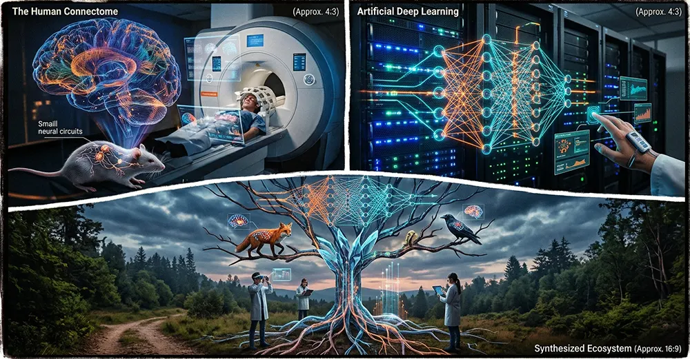
19. A Shared History Between Biology, Computer Science and Artificial Intelligence
The history of the nervous system and the history of artificial intelligence belong to two distinct scientific fields.
Yet they meet around a common question: how can a collection of elementary units process information and produce complex behaviours?
Biology has gradually uncovered the mechanisms through which neurons communicate, organise themselves and modify their connections.
Mathematics and computer science subsequently developed models capable of representing certain general properties of these systems.
This encounter gave rise to artificial neural networks, followed by deep learning and a substantial part of contemporary artificial intelligence.
The relationship between these two fields is not, however, simply a transfer of knowledge from the brain to the machine.
It has become reciprocal.
Artificial models make it possible to test certain hypotheses about the organisation of information.
Neuroscience provides observations that inspire new computer architectures.
Advances in imaging and machine learning make it possible to map neural circuits whose study was previously beyond technical reach.
This convergence opens up considerable prospects.
It concerns, in particular, autonomous robotics, brain-computer interfaces, neuromorphic computing, computational neuroscience and the understanding of learning mechanisms.
Conclusion: From the First Neural Network to Artificial Intelligence, a Shared Question About Information Processing
The history of the nervous system tells the story of a fundamental transformation in the organisation of living systems.
Long before the emergence of the human brain, organisms had developed mechanisms capable of detecting their environment, transmitting information and producing appropriate responses.
The appearance of neurons made it possible to specialise these functions within multicellular organisms.
Diffuse neural networks, ganglia, nerve cords and the centralised systems of vertebrates bear witness to the diversity of architectures developed throughout evolution.
In human beings, this organisation has reached a remarkable degree of complexity.
Neurons, synapses, glial cells and the various brain structures form a dynamic system capable of simultaneously regulating vital functions, perception, movement, memory and cognitive activity.
The brain, however, is not a machine composed solely of cells transmitting electrical impulses.
It is an integrated biological system whose properties emerge from interactions between electrical, chemical, metabolic, cellular and behavioural mechanisms.
It is precisely this complexity that explains the considerable interest it has generated in the field of artificial intelligence.
Artificial neural networks have adopted certain general principles of neuronal functioning to build systems capable of learning from data.
They have gradually evolved towards multilayer architectures, convolutional networks, recurrent models and Transformers, which today constitute major artificial intelligence technologies.
But artificial intelligence is not a digital brain.
It represents another way of conceiving information processing, based on mathematics, computer science and engineering.
The difference between the two systems does nothing to diminish the richness of their scientific interactions.
On the contrary, it is one of the driving forces behind contemporary research.
Neuroscience makes it possible to discover the principles underlying the organisation of living systems. Computer science makes it possible to construct models capable of reproducing certain functional properties. Artificial intelligence, in turn, contributes to analysing biological data and exploring new hypotheses.
From the first cell capable of responding to its environment to contemporary computer architectures, one question thus spans several billion years of evolution and several centuries of scientific research: how can matter organise itself so as to receive, transform, store and use information?
This question constitutes one of the major meeting grounds of biology, neuroscience, mathematics and artificial intelligence.
Cet article est rédigé par Blaise Grenier, Journaliste reporter d'images et auteur. Découvrir le parcours de l'auteur et la charte éditoriale »